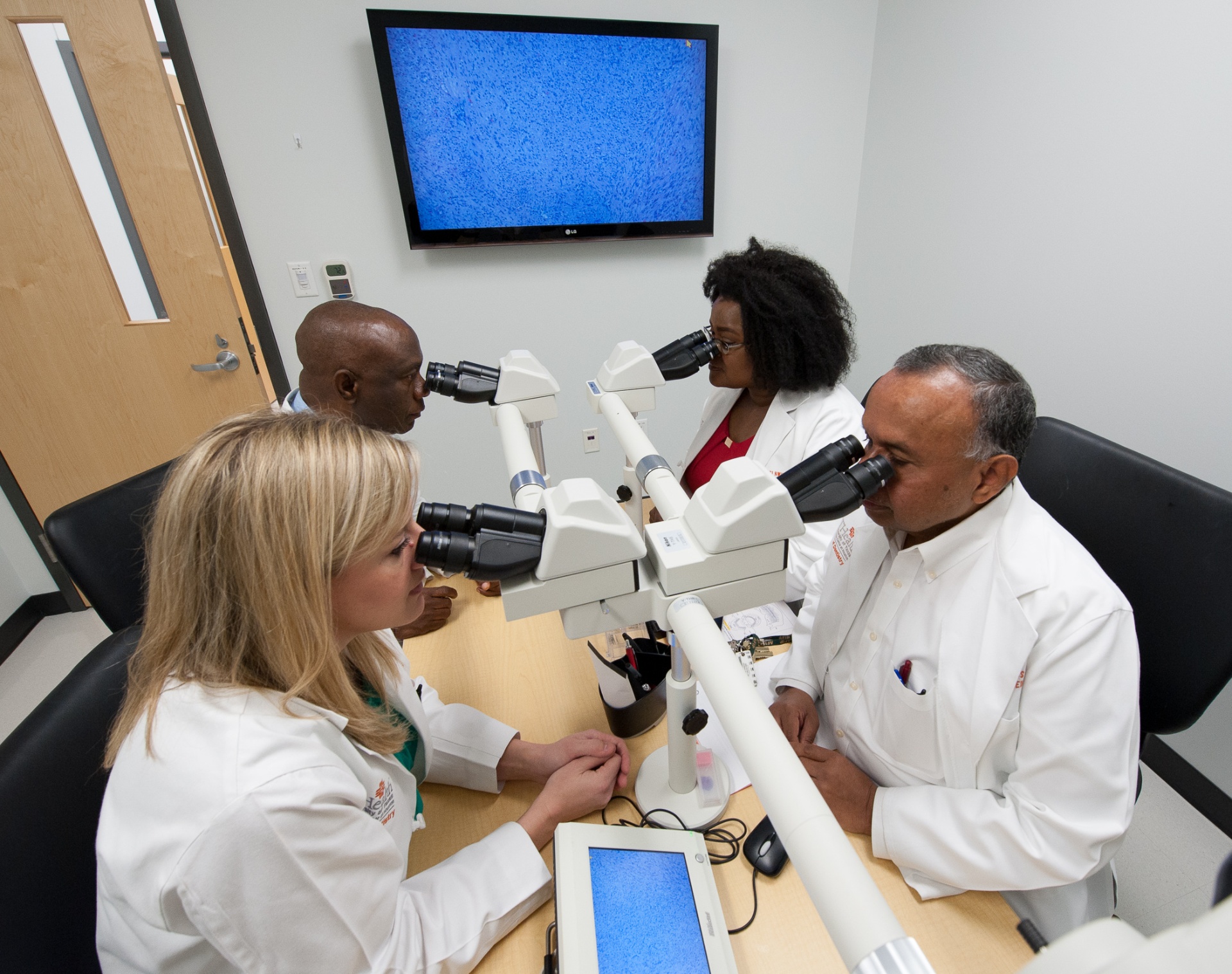
Biopsy Kits
Biopsy Kit Information
Are you a doctor needing to send in a patient’s biopsy specimen for pathology services? Please review this information for answers to frequently asked questions.
- What is the cost of a biopsy kit?
Biopsy kits are supplied free of charge. Request a biopsy kit
- What does a biopsy kit contain?
- Specimen container with 10% Neutral Buffered Formalin
- Requisition form (front and back should be completed)
- Billing information sheet for your patient
- Pre-payment instructions for patients self paying
- ABN instructional sheet
- ABN form needed for Medicare or Medicare patients (MUST be filled out if patient has Medicare OR is of Medicare age — 65 or older)
- Prepaid 2-day FedEx air bill to return the specimen kit to us
*If a specimen for Immunofluorescence is being taken, please refer to the instructions on this webpage on how to collect and submit this type of specimen. Michel’s solution for specimens that are being collected for immunofluorescent testing is supplied free of charge upon request.
- How are biopsy reports communicated?
Biopsy reports are communicated to the surgeon via an electronic reporting system called eDossea, which is a third party, HIPAA compliant platform. Biopsy reports are encrypted to ensure patient privacy.
Biopsy reports can be accessed by the contributing surgeon using a passcode chosen by the contributor that is set up in the system. This same passcode will need to be entered for each report that is sent. If more than three attempts are made to try and open the report, or the report has been opened three times, the system will not allow you to open it again. The laboratory must send it again to the contributing office.
Biopsy reports are NOT sent directly to patients. It is the responsibility of the contributing dentist/surgeon receiving the report to share the report with the patient. Any requests for patient reports to be sent to anyone other than the contributing dentist/surgeon must have a patient signed Authorization for the Use and Disclosure of PHI form for release. To obtain this form contact the lab.
- How do I ship the specimen to the laboratory?
Biopsy kits are shipped to contributing dentist’s/surgeon’s offices via FedEx or USPS. Once the biopsy has been taken and placed in the specimen container provided in the kit, follow these steps:
- Place the specimen container in the plastic biohazard bag containing the absorbent pad. Do NOT place absorbent pad inside the formalin bottle. Ensure the lid clicks in place on the specimen container so the lid is properly secured.
- Ensure specimen bottle is labeled with all requested information: Patient Name, Date of Birth, Date of Procedure, Location of Biopsy, and Doctor’s Name
- Place the sealed specimen bag along with ALL necessary documentation into the biopsy kit
- Secure the biopsy kit shut with a small piece of tape
- Tape the provided prepaid 2-day FedEx air-bill to the outside of the biopsy kit
- Record and keep the tracking number on the air-bill in case the package needs to be tracked while in route to our laboratory
- Call FedEx at 1-800-463-3339 and inform them that you have a package for pick up that has a pre-paid air-bill
- Have a kit, but need a pre-paid FedEx air-bill?
Complete a FedEx Air Bill Request Form or call, fax or email us.
Contact Us
Phone: 713-486-4407
Fax: 713-486-0415
E-mail: [email protected]
Direct immunofluorescence (DIF)
Direct immunofluorescence studies are performed on specimens where additional testing is needed for a definitive diagnosis of a mucocutaneous disorder such as lichen planus, pemphigoid, pemphigus, or erythema multiforme.
IMPORTANT NOTE: Immunofluorescence testing requires the submission of specimens in formalin as well as Michel’s solution in order to obtain the best diagnostic outcome possible. Michel’s solution is available free of charge, upon request.
DIF Specimen Collection Guidelines
- Specimens taken for DIF of bullous/desquamative lesions must be accompanied by another specimen for routine histopathology.
- One specimen goes into formalin, the other into special DIF transport media (Michel’s solution) supplied on request from our lab.
The diagnosis of bullous/desquamative lesions requires an assessment of where the epithelium is splitting. Clinically, obtaining these biopsies can be challenging due to the tendency for the epithelial layer to slough. It is best to seek a site in the anterior portion of the mouth and take care not to grasp the specimen with forceps. Eight-millimeter punch biopsies work quite well, and one should always biopsy the edge of the lesion to include both potentially pathologic and normal appearing tissues..
Immunoglobulins from rabbits, goats or mice are used as reagents. These immunoglobulins are directed to antigens in the host tissue and many of these antigens are immunoglobulins or autoantibodies (i.e. an antibody to an antibody). Many antigens are damaged by formalin fixation and tissue processing; therefore a special medium is used and the sections are cut from frozen tissue samples.
The anti-human immunoglobulins are tagged with a fluorescent dye that can only be visualized with an ultraviolet light source (fluorescent) microscope.
- Localization of fluorescent antibodies
- Basement membrane fibrinogen: lichen planus, lichenoid lesions
- Basement membrane C-3, IgG, IgM and/or IgA: mucous membrane pemphigoid
- Basement membrane IgM: lupus erythematosus
- Intercellular (desmosomal region) C-3, IgG, IgM and/or IgA: Pemphigus vulgaris
- Vascular wall C-3 and IgG: Erythema multiform
Request for Michel’s Solution Biopsy Kit
For collecting the DIF specimen there needs to be both a formalin specimen and a specimen in Michel’s solution submitted.
